Comparative in vitro study of apically extruded material after different technique of root canal instrumentation
Root
canal instrumentation - apically extruded material
Luis
Pascoal VANSAN
Jesus
Djalma PÉCORA
Wanderley
Ferreira da COSTA
Ricardo
Gariba SILVA
Ricardo
Novak SAVIOLI
Este
trabalho está publicado no Brazilian
Dental Journal 8(2(:79-83, 1997
Disciplina
de Endodontia da Faculdade de Odontologia de Ribeirão Preto - USP,
Ribeirão Preto, SP, Brasil.
SUMMARY
Forty
extracted human upper central incisors were submitted to root canal instrumentation
1 mm from the apex by the standard, step-preparation, crown-down, and ultrasound
techniques, using distilled and deionized water as irrigating solution.
The
extrusion product was collected into a collecting device for extruded material
especially fabricated for this purpose. Extrusion was calculated by the
determination of the mass of extruded material.
The
step-preparation technique promoted a larger amount of extrusion than the
standard technique, which in turn promoted greater extrusion than the crown-down
and ultrasound techniques. All techniques used promoted extrusion of material
beyond the apical foramen.
Key
Words: Root canal instrumentation, apically
extruded material.
Introduction
A
successful intervention in the root canal consists of the cure and repair
of the tissues involved and is based on the fulfillment of the following
requirements: chemical-mechanical instrumentation, microbiological control
and root canal sealing. It should be pointed out that the various phases
of endodontic treatment are interdependent, with equivalent and additive
importance and responsibility in terms of successful or unsuccessful total
treatment. Among these phases, chemical-mechanical instrumentation of the
root canal is the one requiring most time and best preparation on the part
of the professional.
Several
studies have shown that dentin filings, necrotic tissue, pulp remnants,
microorganisms and irrigating solution may be forced towards the periapical
tissues during root canal instrumentation. Thus, extrusion of the material
is a problem that occurs with the use of various instrumentation techniques
(Heuer, 1963; Chapman et al, 1968; Vandevisse & Brilliant, 1975; Martin
& Cunningham, 1982; Fairbourn et al, 1987; Ruiz-Hubard et al, 1987;
McKendry, 1990; Lee et al, 1991).
The
objective of the present study was to compare the quantity of material
extruded through the apical foramen during root canal instrumentation using
four different instrumentation techniques, i.e., standard, step-preparation,
crown-down and ultrasound.
Material
and Method
Forty
newly extracted human upper central incisors were used. The teeth had a
fully formed foramen and were of approximately equal length, i.e., 21 to
22 mm, and their apical foramen had a diameter similar to that of a 20
file. For the three techniques of manual instrumentation we used 15 to
40 K Maillefer files, and for ultrasound we used an Enac apparatus coupled
to a pressurized reservoir containing the irrigating solution. Mani K files
were used with the ultrasound apparatus.
Distilled
and deionized water was used as irrigating solution for all techniques
studied, in the volumes routinely applied in each technique.
Four
groups of 10 teeth each were used for each instrumentation technique. All
instrumentation techniques were executed with the tooth fixed in a device
for the collection of extruded material previously fabricated for this
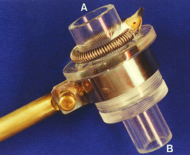
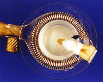
study (Figure 1D). The device consists of an acrylic body with a lateral
orifice at an angle that permits placement of the teeth attached to a spring
always at the same inclination (Figure 1E). The device contains an opening
in the upper end that permits contact with the root of the tooth and another
opening in the lower end for drainage of the irrigating solution utilized.
This lower end is removable and is fitted with a screw-on adaptation measuring
20 mm in diameter (Figure 1A) used to hold the filter paper (Figure 1B).
VER ANEXO I
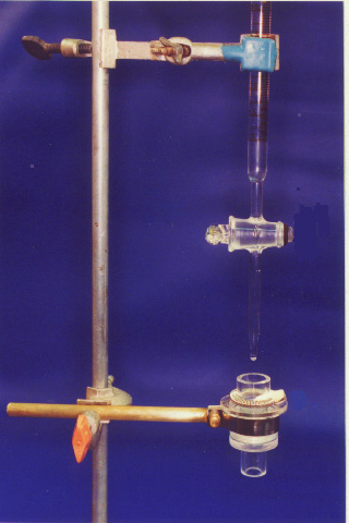
This
ensemble is connected to a metal rod with a clamp and fixed to a universal
support fitted with two metal clamps, one of them used to fix the metal
rod and the other used to hold a 25 mm burette containing distilled and
deionized water (Figure 1 C). The objective was to wash the root portion
at the end of each instrumentation, spilling onto the filter paper the
remaining extrusion material, which adhered to the outer portion of the
apical third.
A
pilot study showed that filter paper disks moistened with 3 ml distilled
and deionized water would dry and reacquire the initial mass after being
placed in an oven at 37oC for 90 minutes.
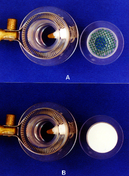
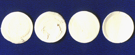
Extrusion
was calculated by weighing the materials that passed through the apical
foramen of the tooth and were collected on the filter paper disk, as shown
in figure 1 F1 for standard instrumentation, in figure 1 F2 for step-preparation,
in figure 1 F3 for the crown-down technique, and in figure 1 F4 in the
ultrasound technique. As a safety margin, we determined that the disks
containing the extruded material needed to be left in the oven at 37oC
for 3 hours. The weight was considered to be final only when it maintained
the same value after three consecutive weight determinations made at 30-minute
intervals.
The
mass of the extruded material was calculated as the difference between
the mass of the paper disk before and after collection of the material
extruded through the foramen opening of the tooth during root canal instrumentation.
All
weight determinations were performed with an MLW scale with a margin of
error of 0.01 mg.
Results
The
experimental data of the present study consisted of 40 numerical values
corresponding to the mass (mg) of the materials extruded through the apical
foramen as a function of the techniques for root canal instrumentation
utilized. The values were obtained from the factorial product of 4 instrumentation
techniques x 10 teeth (replications) (table I).
TABLE
I Mass (mg) of the material extruded through the apical foramen of the
root canal.
| |
Techniques
for root canal instrumentation |
| Replications |
Standard |
Step-Preparation |
Crown-Down |
Ultrasound |
| 1 |
19.8 |
26.6 |
0.4 |
1.2 |
| 2 |
18.5 |
49.1 |
1.1 |
0.6 |
| 3 |
15.5 |
48.6 |
4.8 |
1.8 |
| 4 |
17.6 |
33.0 |
0.8 |
2.6 |
| 5 |
9.6 |
40.4 |
0.4 |
1.4 |
| 6 |
26.5 |
33.1 |
1.0 |
1.9 |
| 7 |
24.5 |
25.6 |
5.9 |
3.4 |
| 8 |
22.8 |
38.4 |
5.0 |
2.3 |
| 9 |
23.5 |
18.3 |
3.5 |
2.1 |
| 10 |
9.5 |
50.1 |
2.5 |
3.0 |
Preliminary
statistical tests applied to the original data indicated that data distribution
was not normal. The data were then transformed to square roots in order
to improve the unfavorable characteristic of sample distribution. When
preliminary tests were again applied to determine normality, it was concluded
that when the square roots of the original data were used the sample distribution
was normal and homogeneous, thus permitting the application of parametric
statistical tests. Analysis of variance showed high significance at the
1% level of probability for the hypothesis of equality, indicating that
there were differences between the techniques for root canal instrumentation
studied in terms of their action in promoting extrusion of the material
through the apical foramen. A complementary Tukey test was carried out
to compare the mean values for the extrusions provoked by the four techniques
(table II).
TABLE
II Tukey test comparing the mean values for the four techniques (square
roots of the data)
| Technique |
Means |
Critical
value (a = 0.1) |
| Step-preparation |
5.96# |
|
| Standard |
4.27+ |
1.05 |
| Crown-down |
1.45* |
|
| Ultrasound |
1.39* |
|
Means
followed by equal symbols did not differ significantly
Discussion
Root
canal instrumentation requires technical knowledge to be applied to the
biological area, so as to obtain a well instrumented and disinfected canal
without damage to its biological structure. Since the root canal includes
the space that contains the pulpar organ, one of its ends is in the pulp
chamber and the other(s) correspond to the apical foramina. Thus, the act
of instrumentation of the root canals, of itself, causes the possible extrusion
of material through the foramen by virtue of the anatomy of the canal itself.
Researchers
have long been trying to develop instrumentation techniques that will minimize
this problem. In the present study we made an attempt to develop a specific
experimental model using natural teeth in which all possible variables
could be controlled, with only the extrusion in the presence of the different
techniques of root canal instrumentation being left for concrete analysis.
Root
canal instrumentation using filing movements may force the material to
the periapex since, as pointed out by Grossman (1956), files act as pistons.
The present findings confirm these observations, as shown by the high index
of material extrusion observed with the conventional and step-preparation
techniques compared to the ultrasound and crown-down techniques, in which
the canal is instrumented with vibratory and rotary movements, respectively.
The step-preparation technique promoted more extrusion probably because,
in addition to the use of the standard technique for the apical third,
the memory instrument that runs along the entire length of the canal is
employed after each instrument of larger caliber is used when backing up
during root canal instrumentation, a fact that can cause extrusion.
The
present results also agree with those reported by Ruiz-Hubard (1987) who
demonstrated a better performance of the crown-down technique compared
to the step-preparation technique. The present results concerning the amount
of material extruded with the use of the ultrasound technique agree with
those reported by Martin & Cunningham (1982).
The
present observations showing that all instrumentation techniques used in
the study promoted extrusion of the material beyond the apical foramen
confirm and support the data reported by other investigators, who stated
that extrusion is unavoidable with the use of any instrumentation technique
(Chapman et al, 1968; Weine, 1976; Martin & Cunningham; 1982, Fairbourn
Et Al, 1987; Ruiz-Hubard et al, 1987; Mckendry, 1990; Lee et al, 1991;
Myers & Montgomery, 1991).
A
general overview of the present results shows that they almost fully agree
with those reported in the literature.
Conclusions
On
the basis of the methodology employed and of the results obtained, we conclude
that:
1.
there was extrusion of material through the apical foramen of the teeth
with the use of all techniques of root canal instrumentation studied.
2.
The step-preparation technique promoted greater extrusion of material through
the apical foramen than the other techniques studied.
3.
The ultrasound and crown-down techniques form a pair, with statistically
similar results and promoted less extrusion of material through the apical
foramen.
4.
The standard technique promoted less extrusion of material through the
apical foramen than the step-preparation technique and greater extrusion
than the crown-down and ultrasound techniques.
References.
Chapman
CE, Colle JG, Beagrie GS: A preliminary report on the correlation between
apical infection and instrumentation in endodontics. J Br Endod Soc 2:
7-11, 1968
Fairbourn
DR, McWalter GM, Montgomery S: The effect of four preparation techniques
on the amount of apically extruded debris. J Endod 13: 102-8, 1987
Heuer
MA: The biomechanics of endodontic therapy. Dent Clin North Am 13: 341-59,
1963
Grossman
LI: Tratamento dos canais radiculares. 2nd ed. Rio de Janeiro, Atheneu
1956.
Lee
SJ, Strittmatter EJ, Lee CS: A compararison of root canal content extrusion
using ultrasonic and hand instrumentation. Endod Dent Traumatol 7: 65-8,
1991
Martin
H, Cunningham WT: The effect of endosonic and hand manipulation on the
amount of root canal material extruded. Oral Surg 53: 611-3, 1982
McKendry
DJ: Comparison of balanced forcs endosonic, and step-back filling instrumentation
technics: quantification of extruded apical debris. J Endod 16: 24-7, 1990
Myers
GL & Montgomery S: A comparison of weights of debris extruded apically
by conventional filing hand canal master thecniques. Endod 17: 275-9, 1991
Ruiz-Hubard
EE, Gutman JL, Wagner MJ: A quantative assessiment of canal debris forced
periapically during root canal instrumentation using two different techniques.
J Endod 13: 554-8, 1987
Weine
F S, Kelly R F, Bray K E: Effect of preparation with endodontic hand piece
on original canal shape. J Endod 2: 298, 1976
Vande-Visse
JE, Brilliant JD: Effect of irrigation on the production of extruded material
at the root apex during instrumentation. J Endod 1: 243-6, 1975
COPYRIGHT
1999 Update
21/sept, 1999
Esta
página foi elaborada com apoio do Programa Incentivo à Produção
de Material Didático do SIAE - Pró - Reitorias de Graduação
e Pós-Graduação da Universidade de São Paulo
ANEXO I
| Figure 1 |
| Figure 1 Device constructed to collect extruded material. |
| Figure 4 |
Figure 5 |
| Figure 4. |
Figure 5. Paper discs showing collected material
after instrumentation. |
|


