Evaluation of Er:YAG Laser and EDTAC on Dentin Adhesion of Six Endodontic Sealers
Authors:
Jesus Djalma PÉCORA1
Antônio Luís CUSSIOLI1
Danilo M. Zanello GUERISOLI1
Melissa A. MARCHESAN1
Manoel D. SOUSA-NETO2
Aldo BRUGNERA-JUNIOR31Faculty of Dentistry of Ribeirão Preto, Universityof São Paulo, Ribeirão Preto, SP, Brazil
2Faculty of Dentistry, University of RibeirãoPreto (UNAERP), Ribeirão Preto, SP, Brazil
3University Camilo Castelo Branco, Dental Associationof the State of São Paulo, São Paulo, SP, Brazil
Correspondence: Prof. Dr. Jesus Djalma Pécora, Departamento deOdontologia Restauradora, Faculdade de Odontologia de Ribeirão Preto,Universidade de São Paulo, 14040-904 Ribeirão Preto, SP,Brasil. e-mail: pecora@forp.usp.br
Braz Dent J (2001) 12(1): 27-30 ISSN 0103-6440 INTRODUCTION | MATERIALAND METHODS | RESULTS | DISCUSSION| RESUMO | REFERENCES
The effect of Er:YAG laser application and EDTAC on the adhesion ofepoxy resin-based endodontic sealers to human dentin was evaluated invitro. A total of 99 extracted human maxillary molars with their crownsworn flat were used. The teeth were divided into 3 groups: group 1, thedentin surface received no treatment; group 2, EDTAC was applied to thedentin surface for 5 min; group 3, the dentin surface received Er:YAG laserapplication (2.25 W potency; 11 mm focal distance; 4 Hz frequency; 200mJ energy; 62 J total energy; 313 mean impulse). Three teeth from eachgroup were analyzed by scanning electron microscopy for changes in dentinsurface. The epoxy resin root canal sealers used were: AH Plus®,Topseal®, Sealer 26®, AH 26®,and Sealer Plus®. The zinc oxide eugenol-based sealer Fillcanal®was used as control. Adhesion was measured with a Universal testing machine.The results showed a statistically significant difference at the levelof 1% among the dentin treatments. The dentin treated with Er:YAG lasershowed greater adhesion with the sealers than dentin treated with EDTACwhich was greater than dentin that received no treatment. The Tukey testshowed the formation of 5 groups in decreasing order of adhesion: AH Plus,Topseal and Sealer 26, AH 26, Sealer Plus, and Fillcanal (Grossman cement). Key words: Endodontics, Er:YAG laser, root canal sealer.
INTRODUCTION Because a hermetic seal of root canals is essential in Endodontics,sealing materials and their properties are of vital importance for thesuccess of root canal treatment. The properties of the root canal sealerscan be classified as physico-chemical, antimicrobial and biological. In1984, a series of regulations and tests were made effective, created oneyear earlier by the American Dental Association (1) for the standardizationof research of these properties. However, no model for adhesion and apicalmicroleakage tests was adopted. Adhesion of a root canal sealer means its capacity to attach to thedentinal walls of the root canal and provide bonding between it and gutta-perchapoints. Ørstavik (2) reported the use of the Universal testing machineto perform adhesion tests of the root canal sealers. This method was alsoused by Hyde (3), Wennberg and Ørstavik (4), and Sousa-Neto (5). During the chemo-mechanical preparation of the root canal, a smear layeris produced, which is a negative factor in root canal obturation, becauseit is constituted by organic and inorganic material located on the interfacebetween the root canal walls and the sealing material and weakly attachedto them. Thus, it interferes in the adhesion of the sealing material tothe root canal walls (6-8). Researchers have recommended a final flushof EDTA (ethylenediaminetetraacetic acid) after the biomechanical preparationof the root canal in order to remove this smear layer (9-11). Recently,Takeda et al. (12) showed the capacity of Er:YAG laser in removing smearlayer. Sousa-Neto (13) reported that after Er:YAG application on dentinsurface, the adhesion of Sealer 26 increased. The objective of the present research is to evaluate the effect of EDTACand Er:YAG laser on the adhesion of epoxy resin-based root canal sealersto human dentin.
MATERIAL AND METHODS The enamel of the occlusal surface was removed from ninety-nine humanmolars from laboratory stock with a number 4138 diamond bur (KG Sorensen,Barueri, SP, Brazil) in a high speed handpiece cooled with water. Afterobtaining a flat occlusal surface, the teeth were fixed by their rootsin an acrylic resin base to be adapted to the Instron Universal testingmachine (model MEM 2000, Curitiba, PR, Brazil). The teeth were divided randomly into 3 groups of 33 samples each. Thefirst group received no treatment at all. In the second group, 50 µlof EDTAC was placed on the dentin surface for 5 min. This EDTAC solutionwas fabricated at the Endodontics Research Laboratory (FORP-USP, RibeirãoPreto, SP, Brazil) and consists of an aqueous solution of 15% EDTA (Merck,Rio de Janeiro, RJ, Brazil) at pH 7.3 and 0.1% Cetavlon®(cetyltrimethylammonium) (Sigma, St. Louis, MO, USA). The third group receivedirradiation with Er:YAG laser (KaVo Key laser II, Warthausen, Germany)on the dentin surface, with the following parameters: 11 millimeters focaldistance perpendicular to the dentin surface, 4 Hz frequency, 200 mJ energy,1 min application time and 2.25 W power, totaling 62 J of energy. Six root canal sealers were used in this experiment: Sealer 26 (Dentsply,Petrópolis, RJ, Brazil), AH 26 (Dentsply, Konstanz, Germany), AHPlus (De Trey-Dentsply, Konstanz, Germany), Sealer Plus (Dentsply, Petrópolis,RJ, Brazil), Topseal (Dentsply-Maillefer,
Ballaigues, Switzerland) and Fillcanal (D.G. Ligas Odontológicas,Rio de Janeiro, RJ, Brazil). Five repetitions for each experimental groupwere performed for each root canal sealer. Aluminum cylinders (10 mm high x 6 mm internal diameter) with stainlesssteel handles were placed on the dentin surface and fixed laterally tothe teeth with wax. After preparation according to manufacturers' directions,the sealers were carefully placed inside the cylinders with the aid ofa vibrator. The material was then placed at 37oC, 95% humidityfor a period 3 times greater than the normal setting time of the sealer. The samples were placed in an Instron Universal testing machine (modelMEM 2000), with a grip that held the aluminum cylinder handle and exertedvertical traction. The machine was calibrated to run at a constant speedof 1 mm/min until the aluminum cylinder containing the sealer was detached.The traction force necessary to detach the device from the tooth, givenin kilograms-force (KgF) and later transformed to MegaPascal (MPa), wasrecorded. The Tukey test was used for statistical analysis (p<0.01).Three teeth from each group were evaluated by scanning electron microscopyfor changes in the dentin surface.
RESULTS 
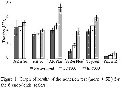
Figure 1 shows the mean values of the adhesiontest for the 6 sealers. Statistical analysis showed significant differences(p<0.01) between the tested sealers, except for Topseal and Sealer 26,that were statistically similar. The sealers can be ranked in decreasingvalues of adhesion to dentin: AH Plus, Sealer 26 and Topseal, AH 26, SealerPlus, and Fillcanal. The treatments for dentin showed significant differences (p<0.01)between treatments, with higher adhesion values for dentin irradiated withEr:YAG laser. Dentin with no treatment revealed the lowest adhesion values,and dentin treated with EDTAC was in an intermediate position.
DISCUSSION Adhesion is among the properties that an ideal root canal sealer musthave, according to Grossman (14) and Branstetter and Fraunhofer (15). The American Dental Association, in 1983, established a series of regulationsand tests for the studyof the physical properties of root canal sealers.However, due to the lack of consensus among researchers, adhesion testswere not standardized. Ørstavik (2) proposed the use of the Universaltesting machine to test root canal sealer adhesion. This method was alsoused by Hyde (3), Wennberg and Ørstavik (4) and Sousa-Neto (5,16).The Universal testing machine promotes better uniformity and greater reproducibility,providing more accurate results, and tension values in MegaPascal (MPa)favor comparison of results, because it is an internationally acceptedunit. The adhesion of Grossman type root canal sealers to dentin is establishedby electrostatic bonding and not by its penetration into the dentinal tubules.For this reason, the action of EDTAC and Er:YAG laser did not significantlyincrease adhesion values of Fillcanal to dentin. Ionic bonding betweenrosin and dentin is weak by its own nature (5). Epoxy-based sealers show higher adhesion to dentin and among these,AH Plus had the highest values for the traction test. Topseal and Sealer26 presented inferior adhesion values compared to AH Plus, but superiorto the other tested sealers. According to the manufacturers (Dentsply),Topseal and AH Plus have the same formulation. The difference between themlays in the fact that AH Plus is produced by De Trey, a Dentsply subsidiaryin Germany, and Topseal is produced by Dentsply-Maillefer in Switzerland.Manufacturers in different countries use chemical compounds acquired andproduced in different places with different manufacturers and packing.In terms of adhesion to dentin, they have distinct behaviors. Sealer 26,produced by Dentsply from Brazil, showed adhesion to dentin similar toTopseal. It is an epoxy-based sealer with a formula similar to AH SilverFree, except that Sealer 26 has calcium hydroxide in its formulation whileAH Silver Free does not. This addition of calcium hydroxide to the formulaimplies a reduction of the bismuth trioxide percentage. 
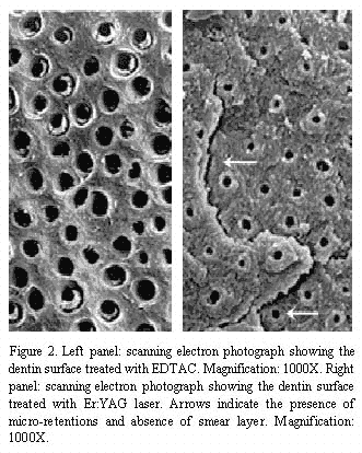
The chelating agent EDTAC increased adhesion values when compared tothe dentin without any treatment. EDTAC removes smear layer, which canpermit the penetration of epoxy-based sealers into the dentinal tubules.This favors a greater bonding between dentin and sealer, increasing theadhesion values compared to dentin without treatment (Figure2). Application of Er:YAG laser to dentin surface caused higher adhesionof epoxy-based sealers. This
can be explained by two factors: a) the removal of smear layer, exposingdentinal tubules that are partially filled with sealer, creates a "tag"(17), and b) the conditioning of the dentin surface, causing a higher mechanicalbonding between sealer and intertubular dentin by increasing the area termedmicro-retentive pattern (18) (Figure 3). This pattern did not increasethe adhesion values of Fillcanal, a zinc oxide eugenol-based sealer. Sousa-Neto (13) reported that application of Er:YAG laser to dentinpromoted an increase in adhesion values of Sealer 26. This was confirmedby this experiment which showed that application of Er:YAG laser and EDTACto the surface of human dentin increased the adhesion of endodontic sealers.It is important to remember that the laser beam was applied perpendicularlyto the dentin surface and that the results obtained may be due to thisdirection. Within root canals, the laser beam is applied with an opticfiber and the beam direction may be different. This factor needs to beevaluated further. Thus, this study opens perspectives for more research to investigatethe effect of direction of the laser beam when applied within the rootcanal using various diameters of optic fiber and also if greater adhesionleads to less marginal infiltration.
ACKNOWLEDGMENTS Research was supported by a grant from CNPq to Dr. Jesus D. Pécora.
RESUMO Pécora JD, Cussioli AL, Guerisoli DMZ, Marchesan MA, Sousa-NetoMD, Brugnera-Junior A. Estudo in vitro do efeito da aplicaçãodo laser Er:YAG e da solução de EDTAC na superfíciedentinária sobre a adesividade de seis cimentos endodônticos.Braz Dent J 12(1):27-30. Estudou-se in vitro o efeito da aplicação do laserEr:YAG e da solução de EDTAC na superfície dentináriasobre a adesividade de diferentes cimentos endodônticos àbase de resina epóxica. Foram utilizados 99 molares superiores humanosde estoque que tiveram suas coroas desgastadas até se obter umasuperfície plana transversal ao longo eixo do dente e foram divididosem três grupos com 33 dentes cada. No primeiro grupo, a superfíciede dentina não recebeu nenhum tratamento. No segundo, aplicou-sesobre a dentina uma solução de EDTAC por cinco minutos e,no terceiro, a dentina recebeu a aplicação do laser Er:YAGcom os seguintes parâmetros: potência 2,25 W; distânciafocal 11 mm; freqüência de 4 Hz; período de aplicaçãode 1 minuto e energia de 200 mJ, totalizando 62 J de energia aplicadosao dente. Três dentes de cada grupo foram enviados para a análisede microscopia eletrônica de varredura. Os cimentos endodônticosà base de resina epóxica testados foram os seguintes: AHPlus, Topseal, Sealer 26, AH 26 e o Sealer Plus. O cimento Fillcanal, cimentotipo Grossman à base de óxido de zinco e eugenol, foi utilizadocomo controle. A força de adesão foi detectada por meio deuma Máquina Universal de Ensaios. Os resultados evidenciaram haverdiferença estatística significante ao nível de 1%de probabilidade entre as condições de tratamento da dentinae os diferentes cimentos endodônticos. Assim, a dentina tratada comlaser Er:YAG propiciou a maior adesividade, a dentina tratada com a soluçãode EDTAC proporcionou adesividade intermediária e a dentina quenão recebeu tratamento algum mostrou a menor adesividade. No quediz respeito aos cimentos endodônticos testados, o teste de Tukeymostrou a formação de 5 grupos em ordem decrescente de adesividadeà dentina: AH Plus, com a maior adesividade; Topseal e Sealer 26,com valores estatisticamente semelhantes; AH 26; Sealer Plus; e Fillcanal,com o menor valor de adesividade. Unitermos: endodontia, laser Er:YAG, cimentos obturadores dos canaisradiculares.
REFERENCES 1. American National Standards Institute Specification 57 for Endodonticfilling materials. J Am Dent Assoc 1994;108:88. 2. Ørstavik D. Weight loss of endodontic sealers, cements andpastes in water. Scand J Dent Res 1983;91:316-319. 3. Hyde DG. Physical properties of root canal sealers containing calciumhydroxide. [Master's thesis]. Michigan: University of Michigan; 1986. 80p. 4. Wennberg A, Ørstavik D. Adhesion of root canal sealers tobovine dentine and gutta-percha. Int Endod J 1990;23:13-19. 5. Sousa-Neto MD. Estudo da influência de diferentes tipos debreus e resinas hidrogenadas sobre as propriedades físico-químicasdo cimento obturador dos canais radiculares do tipo Grossman. [Doctoralthesis]. Ribeirão Preto: Faculdade de Odontologia de RibeirãoPreto, Universidade de São Paulo; 1997. 108 p. 6. White RR, Goldeman M, Peck SL. The influence of the smeared layerupon dentinal tubule penetration by plastic materials. J Endodon 1994;10:558-562. 7. Kennedy W, Walker WA, Gouch RW. Smear layer removal effects on apicalleakage. J Endodon 1986;12:21-27 8. Economides N, Liolios E, Kolokuris I, Beltes P. Long-term evaluationof the influence of smear layer removal on the sealing ability of differentsealers. J Endodon 1999;25:123-125. 9. Yamada RS, Armas A, Goldman M. A scanning electron microscopic comparisonof a high volume final flush with several irrigating solutions: part 3.J Endodon 1983;9:137-142. 10. Garberoglio R, Becce C. Smear layer removal by root canal irrigants.A comparative scanning microscopic study. Oral Surg 1994;78:359-367. 11. Kouvas V, Liolios E, Vassiliadis L, Parissis-Messimeris S, BoutsioukisA. Influence of smear layer on depth of penetration of three endodonticsealers: an SEM study. Endod Dent Traumatol 1998;14:191-195. 12. Takeda FH, Harashima T, Kimura Y, Matsumoto K. A comparative studyof the removal of smear layer by three endodontic irrigants and two typesof laser. Int Endod J 1999;32:32-39. 13. Sousa-Neto MD, Marchesan MA, Pécora JD, Brugnera Junior A,Silva-Sousa YTC, Saquy PC. Effect of Er:YAG laser on adhesion of root canalsealers. J Endodon (in press). 14. Grossman LI. An improved root canal cement. J Am Dent Assoc 1958;56:381-385. 15. Branstetter J, Fraunhofer JA. The physical properties and sealingaction of endodontic sealer cements: a review of the literature. J Endodon1982;8:312-316. 16. Sousa-Neto MD, Guimarães LF, Guerisoli DM, Saquy PC, PécoraJD. Influence of different kinds of rosins and hydrogenated resins on thesetting time of Grossman cements. Rev Odontol USP 1999;13:83-87. 17. Potts T, Pitrou A. Laser photopolymerization of dental materialwith potential endodontics applications. J Endodon 1990;60:265-268. 18. Tanji EY, Matsumoto K, Eduardo CP. Scanning electron microscopicobservations of dentin surface conditioned with the Er:YAG laser. DeutsGesellschaft Laser Newsletter 1997;8:6.
|

