Upper Molar Dens in Dente - Case Report
Wanderley
F. COSTA
Manoel
D. SOUSA NETO
Jesus
D. PECORA
Trabalho publicado
no Brazialian Dental Journal
1(1):45-49, 1990
The
authors present a case report of dens in dente in an upper molar,
in which endodontic treatment was impossible due to its location.
Key
Words: dens in dente, dens invaginatus, therapy.
Introduction
Dens
in dente is an anomaly of development resulting from deepening or
invagination of the enamel organ into the dental papilla, which begins
at the crown and often extends to the root, before the calcification of
the dental tissues.
The
first observation of dens in dente dates back lo 1856 (De Smit and
Demaut, 1982). This anomaly is also known as dens invaginatus, dilated
odontome and gestant anomaly. According to Pécora et al. (1990),
the varied nomenclature occurs probably due to the lack of consensus in
relation to the cause of this alteration and the various names reflect
the different opinions as to its etiology.
Hovland
(1977) calculated the incidence of dens in dente to be 0.04 to 10.00%,
occurring in any tooth but with a greater frequency in the permanent upper
lateral incisors. Cases of bilateral occurrence have been reported (Swanson
and McCarthy, 1947; Grossman, 1976; Burton et al., 1980).
Dens
in dente is classified into 3 types, depending on the depth of invagination.
Type I, the invagination ends in a blind sac, limited to the dental crown.
Type II, the invagination extends to the amelocemental junction, also ending
in a blind sac. Type III, the invagination extends to the interior of the
root, providing an opening to the periodontium, sometimes presenting another
foramen in the apical region of the tooth (Oehlers, 1957).
Radiographically,
dens in dente shows a radiopaque invagination similar in density
to dental enamel (Goaz and While, 1987).
Maisto
(1973), Tagger (1977), Cole et al. (1978), Eldeeb (1984), Pécora
et al. (1987, 1990), and Vajrabhaya (1989) report cases of dens in dentetreated
with conventional endodontic methods. However, Fergunson et al. (1980),
Schindler and Walker (1983), and Pécora et al. (1987, 1990) describe
special endodontic techniques capable of inducing an apexification in Oehlers’
Type II dens in dente. Weine (1982) and Leonardo et al. (1982) report
the endodontic treatment of dens in dente as being a difficult operation,
preferring surgical treatment with retrograde filling.
Case
Report
A
26 year old, white, female patient was referred to our clinic with pain
in the upper left region. Clinical examination showed edema of the vestibular
cul-de-sac region, at the level of the second upper left molar, which did
not respond to thermal tests (heat and cold) but responded positively to
palpation and percussion.
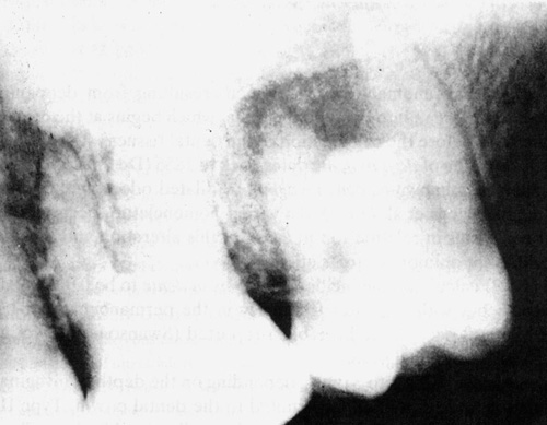
An
x-ray showed the presence of Type III (Oehlers, 1957) dens in denteof the
second upper left molar with an apical radiolucent area (Figure
1).
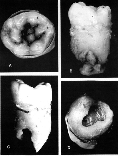
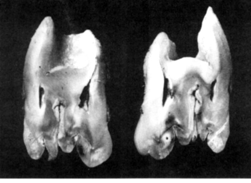
Due
to the localization of the tooth and the impossibility of endodontic root
canal treatment, complemented by the retrograde filling of the apical opening,
the treatment of choice was extraction (Figures
2 and 3).
Discussion
The
presence of dens in dente is more common in the lateral upper incisors,
being rare in molars. Types I and II (Oehlers, 1957) dens in dente do
not present problems in endodontic treatment. In Type II, the invagination
is restricted to the interior of the root canal without reaching the apical
region of the canal, ending in a single foramen.
Pécora
et al. (1987, 1900), Maisto (1973) and Tagger (1977) present methods for
the endodontic treatment of Type II dens in dente.
Hata
and Toda (1987), Bolanos (1988) and Kulild (1989) recommend the endodontic
treatment of dens in dente in anterior upper teeth, even in Type
III (Oehiers, 1957) cases. In these cases, conventional endodontic treatment
in the area of invagination must be complemented by retrograde filling
of the foramen of the principal canal.
In
the case reported here, the first upper left molar presented an apical
radiolucent area and pulp necrosis, probably due to the fact that dens
in dente, Types II and III (Oehlers, 1957), permits the penetration
of irritants into the interior pulp tissue once the invagination establishes
this communication with the buccal cavity. Contamination can also occur
by communication with the pulp through the cul-de-sac of the invagination
causing tissue necrosis.
The
current literature does not present solutions for the treatment of Type
III dens in dente in molars which will only be possible when the
retrograde filling of the root canal is viable.
References
Bolanos
OR, Martell B, Morse DR: A unique approach to the treatment of a tooth
with dens invaginatus. J Endodont 14: 315-318, 1988
Burton
JD, Saffos RO, Scheffer RB: Multiple bilateral dens in dente as
a factor in the etiology of multiple periapical lesions. Oral Surg 49:
496-499,1980
Cole
GM, Taintor JF, James GA: Endodontic therapy of a dilated dens invaginatus.
J
Endodont 4: 89-90, 1978
De
Smit A, Demaut L: Nonsurgical endodontic treatment of invaginatus teeth.
J Endodont 8: 506-511, 1982
Eldeeb
ME: Nonsurgical endodontic therapy of a dens invaginatus. J Endodont
10: 107-109, 1984
Fergunson
FS, Friedman S, Frazzetto V: Successful apexification technique in an immature
tooth with dens in dente. Oral Surg 49: 356-359, 1980
Goaz
PW, White SC: Oral radiology. Principles and interpretation. Mosby, St
Louis 1987
Grossman
LI: Endodontia prática. Guanabara-Koogan, Rio de Janeiro 1976
Hata
I, Toda T: Treatment of dens invaginatus by endodontic treatment
of dens invaginatus, apicocuretage and retro-filling: a case report.
J Endodont 13:469-472, 1987
Hovland
EJ: Nonrecognition and subsequent endodontic treatment of dens invaginatus.
J
Endodont 3: 360-361,1977
Kulild
JC, Weller N: Treatment considerations in dens invaginatus. J Endodont
15: 381-384, 1989
Leonardo
RM, Leal, JM, Simões AP: Endodontia: Tratamento dos canais radiculares.
Panamericana, São Paulo 1982
Maisto
OA: Endodontia. Mundi, Buenos Aires 1973
Oehlers
FA: Dens invaginatus: Variation of the invagination process and
associated anterior crown forms. Oral Surg 10: 1204-1218, 1957
Pécora
JD, Costa WF, Macchetti DD: Caso clínico: Dens in dente. Rev
Odont USP 1: 46-49, 1987
Pécora
JD, Vansan LP, Gariba Silva R, Aiello JSS: Dens invaginatus: Tratamento
endodôntico em uma sessão. Rev Ass Paul Cirurg Dent (in press)
1990
Swanson
WF, McCarthy FM: Bilateral dens in dente. J Dent Res 26: 167-171,
1947
Schindler
WC, Walker WA: Continued root development after apexification of an immature
tooth with dens invaginatus. J Endodont 9: 430-433,1983
Tagger
M: Nonsurgical endodontic therapy of tooth invagination. Oral Surg 43:
124-129, 1977
Vajrabhaya
L: Nonsurgical endodontic treatment of tooth with double dens in dente.J
Endodont 15: 323-327, 1989
Weine
FS: Endodontic therapy. Mosby, St. Louis 1982
Webmasters: J.
D. Pécora e R. S. Silva.
Copyright
2000 Department of Restorative Dentistry
Atualizada
em 25 de janeiro de 2000. Esta página foi elaborada com apoio do
Programa Incentivo à Produção de Material Didático
do SIAE - Pró-Reitorias de Graduação e Pós-Graduação
da USP.
|


