Osseous Regeneration in the Presence of Fibrin Adhesive Material (Tissucol®) and Epsilon-Aminocaproic Acid (EACA)
Tetuo OKAMOTO[1]
Maria Cristina R. ALVES-REZENDE[2]
Ana Cláudia OKAMOTO[1]
Inês A. BUSCARIOLO[1]
Idelmo R. GARCIA Jr.[1]
[1]Disciplina de Cirurgia e Traumatologia Buco-Maxilo-Facial and
[2]Disciplina de Materiais Odontológicos, Faculdade de
Odontologia, UNESP, Araçatuba, SP, Brasil
Braz Dent J (1995) 6(2): 77-83 ISSN 0103-6440
| Introduction | Material/Methods
| Results | One day after surgery
| Three days after surgery | Seven
days after surgery | Fourteen days after
surgery | Twenty-one days after surgery
| Discussion | Conclusions
| References |
The effects of Tissucol® and Tissucol®/EACA on bone healing
were evaluated histologically. Experimental defects were made in both tibias
of 25 rats. Test materials were placed in defects in right tibias and left
tibias served as control. Five animals in each group were killed at 1,
3, 7, 14 and 21 days after surgery. Results showed that: a) Tissucol®
did not interfere with connective and osseous tissue formation; b) Tissucol®
allowed new bone formation; c) Tissucol® residues in Tissucol®
groups in sections of 21-day specimens did not impair healing; d) Tissucol®/EACA
was usually completely resorbed and healing was complete 21 days after
surgery in the Tissucol®/EACA group.
Key words: fibrin sealing, epsilon-aminocaproic acid, bone healing.
Introduction
The effectiveness of hemostatic agents is characterized by their ability
to allow bone regeneration and to undergo resorption.
Studies evaluating the effects of fibrin adhesive material (Tissucol®)
in surgical sites have generally found it to be an effective agent in maintaining
a dry field and allowing new bone formation (Baldin et al., 1985; Palattella
et al., 1985). In addition, results of some experimental (Alves-Rezende
and Okamoto, 1992, 1995; Okamoto et al., 1995) and clinical studies (Matras,
1982; Wepner et al., 1982; Caruso et al., 1984; Stajicic et al., 1985)
suggested that resorption of Tissucol® occurred in the presence of
tissue formation.
Alves-Rezende and Okamoto (1995) reported positive results with the
use of epsilon-aminocaproic acid (EACA) before using the fibrin tissue
adhesive (Tissucol®). They compared Tissucol® and Tissucol®/EACA
as hemostatic agents on alveolar cavity in rats under stress and observed
complete healing in a 24-day period with Tissucol®/EACA. Furthermore,
Tissucol®/EACA showed better resorption than Tissucol® under similar
conditions.
Okamoto et al. (1995) evaluated the effects of Tissucol® on bone
healing in rat tibias and observed small fragments of material 21 days
after implantation. However, they reported that the healing of the experimental
defects was qualitatively similar to those observed in control defects.
Therefore, the present study was designed to evaluate the effects of
fibrin tissue adhesive (Tissucol®) with epsilon-aminocaproic acid (EACA)
on osseous healing in rat tibias.
Material and Methods
Twenty-five male albino (Rattus norvegicus albinus, Wistar) weighing 300
to 350 g were employed. They were fed a diet of standard Purina rat chow
and water ad libitum. At surgery, each animal was anesthetized by intraperitoneal
injection of thionembutal (50 mg/kg body weight). The incision site was
cleaned with 70% ethanol and a 2-3 cm incision was extended distally from
the tibial tubercle. The medial surface of each tibia was exposed and an
opening (one on left tibia and two on right tibia) that extended through
the cortex into the medullary cavity was made in the middle of each surface
5 mm distal to the tibial tubercle with a slow-speed handpiece and # 8
burr. A hemostatic agent was placed in the defects in the tibias of each
animal in the following manner. In the right tibia, Tissucol® (Immuno
A.G., Vienna) was inserted in the upper defect after it was mixed according
to the manufacturer's directions. The lower defect was irrigated with epsilon-aminocaproic
acid (EACA) (5 ml of 5% solution for 2 min) before implanting Tissucol®.
The defect of the left tibia served as an empty control. Incisions were
closed with 4-0 gut sutures placed 3 mm apart. Five animals were killed
by ether overdose at 1, 3, 7, 14 and 21 days after surgery. Appropriate
areas of the tibias were isolated by gross dissection and placed in 10%
formaldeyde. Following fixation, the specimens were decalcified in a 15%
formic acid solution and processed for routine paraffin embedding. Seven-micrometer
sections were cut and stained with hematoxylin and eosin. All sections
were examined with a Zeiss universal microscope.
Results
One day after surgery
The osseous defects were filled with fibrin clot and an influx of inflammatory
cells was seen. In Tissucol® specimens there were neutrophils adjacent
to the test material which occupied a larger portion of the osseous defect.
Some macrophages were observed permeating the fibrin clot, which were more
pronounced in the Tissucol®/EACA specimens and control specimens than
in Tissucol® specimens. In the control sites and Tissucol®/EACA
sites more new budding capillaries and fibroblasts were noted.
Three days after surgery
The osseous defects were filled with an organized fibrin clot which
showed more macrophagic invasion in the Tissucol®/EACA group and control
group than in the Tissucol® group. The test implant was observed in
the center of the defect and there were signs of resorption in Tissucol®/EACA
sites. Fibroblast proliferation was scattered throughout the implant in
Tissucol®/EACA specimens.
Seven days after surgery
Tissucol® and Tissucol®/EACA implant sites showed residual masses
of test material (Figure 1A,B). The implant material was surrounded by
peripheral fibrous connective tissue and there were small amounts of new
bone scattered around the periphery in Tissucol®/EACA specimens. In
the control group, active bone formation was noted throughout the control
defects (Figure 1C).
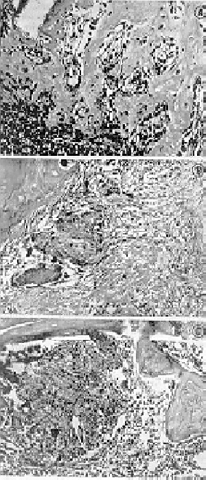
Figure
1 - A, A 7-day Tissucol® specimen demonstrates a residual mass
of material (M) (hematoxylin and eosin, original magnification X160). B,
A 7-day Tissucol®/EACA specimen demonstrates a residual mass of Tissucol®/EACA
(T®/E) surroun-ded by immature bony trabeculae (T) (hematoxylin and
eosin, original magnification X160). C, A 7-day control specimen demonstrates
active bone forma-tion in the center of the osseous defect (hematoxylin
and eosin, original magnification X160).
Fourteen days after surgery
A thicker trabeculae of immature bone occupied most of the defect in
the control group. In the Tissucol® group, occasional material remnants
were observed which were surrounded by immature bony trabeculae or fibrous
connective tissue. In Tissucol®/EACA specimens, a dense trabeculae
of bone was observed in most of the defect. Some specimens showed residual
masses of material while other specimens showed no material.
Twenty-one days after surgery
In the control group, the defect was filled by remodeling compact bone
that was delineated from older bone by cement lines (Figure 2A). Bone regeneration
was complete. In the Tissucol® group, the specimens showed more bone
repair compared with 14-days. Residual masses of material were still present
(Figure 2B). Tissucol®/EACA specimens demonstrated a substantial increase
in the amount of mature bone (Figure 2C). There was no material found in
any of the specimens.
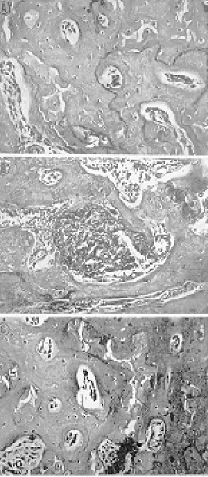
Figure
2 - A, A 21-day control specimen demonstrates cement lines (L) demarcating
newly formed bone (nb) from surround-ing unmanipulated cortical bone (B)
(hematoxylin and eosin, original magnification X160). B, A 21-day Tissucol®
specimen demonstrates a residual mass of the material (M) surrounded by
immautre bony trabeculae (T) (hematoxylin and eosin, original magnification
X160). C, A 21-day Tissucol®/EACA specimen demonstrates the large amount
of dense bone trabeculae filling the experimental defect (hema-toxylin
and eosin, original magnification X160).
Discussion
The healing of control defects in rat tibias was similar to that described
by other investigators (Howard and Kelley, 1969; Ligget et al., 1980; Ibarrolla
et al., 1985; Haasch et al., 1989).
The most significant finding of this study was the beneficial effect
of epsilon-aminocaproic acid (EACA) on resorption of Tissucol®. As
noted in other studies there is a greater probability of resorption of
Tissucol® in the presence of EACA.
In a comparative study of Tissucol® and Tissucol®/EACA, Alves-Rezende
and Okamoto (1995) emphasize the importance of irrigation procedures with
EACA before implant of Tissucol®. We agree that careful irrigation
with EACA before implant of this material is necessary regardless of the
implant site.
Tissucol® did not cause any untoward reactions when placed in bone
defects. Organization and osteogenesis were observed 14 days after implantation,
which is comparable to events observed in controls. Small fragments of
the material were observed in sections of 21-day specimens but did not
cause inflammation or impair healing.
When EACA was used before implant of Tissucol®, it resorbed more
rapidly and there was little resistance to resorption, even when placed
in volumes greater than actually needed to control bleeding at the surgical
sites.
Based on the findings, we believe that Tissucol® and Tissucol®/EACA
should be used as a hemostatic agent in surgical procedures.
Conclusions
In this study, both Tissucol® and Tissucol®/EACA were effective
hemostatic agents. Tissucol® and Tissucol®/EACA did not elicit
foreign body reaction and did not prevent bone healing. The irrigation
procedures with epsilon-aminocaproic acid (EACA) solution before implant
of the Tissucol® facilitated the resorption of the material.
Acknowledgments
This work was supported by FAPESP.
References
Alves-Rezende MCR, Okamoto T: Implante de "Tissucol" em feridas de extração
dental. Estudo histológico em ratos. Rev Odontol UNESP 21: 161-170,
1992
Alves-Rezende MCR, Okamoto T: Effects of fibrin adhesive material (Tissucol®)
on alveolar healing in rats under stress. J Nikon Univ Sch Dent (In press)
Baldin C, Bedeschi G, Beltrame M, Storti E: Sull'impiego di colla di fibrina
umana (Tissucol) in Odontoestomatologia. Giornale di Stomatol e di Ortognat
4: 69-75, 1985
Caruso F, Serpico R, Laino G: Experienze cliniche con la colla di fibrina
in chirurgia paradontale. Arch Stomatol 25: 4: 339-347, 1984
Haasch G, Gerstein H, Austin BP: Effects of two hemostatic agents on
osseous healing. J Endodon 15: 310-314, 1989
Howard IC, Kelley RR: The effect of bone wax on the healing of experimental
rat tibial lesions. Clin Orthop Rel Res 63: 226-232, 1969
Ibarrolla JL, Bjorenson JE, Austin BP, Gerstein H: Osseous reactions
to three hemostatic agents. J Endodon 11: 75-83, 1985
Ligget WR, Brady JM, Tsaknis PJ, Del Rio CE: Light microscopy and microprobe
analysis of bone response to zinc and nonzinc amalgam implants. Oral Surg
49: 254-262, 1980
Matras H: The use of fibrin sealant in oral and maxillofacial surgery.
J Oral Maxillofac Surg 40: 617-622, 1982
Okamoto T, Alves-Rezende MCR, Buscariolo IA, Okamoto AC, Garcia IR:
The effect of fibrin adhesive material (Tissucol) on the healing of experimental
rat tibial lesions. Rev Odont UNESP (In press) Palattella G, Massi C, Corbeli
V, Ruggeri B, Pignatelli N, Palattella E: L'impiego della colla di fibrina
umana liofilizzata "Tissucol" nella chirurgia orale. Dental Cadmos 53:
65-68, 1985
Stajicic Z, Todorovic LJ, Petrovic V: Tissucol in closure of oroantral
communication. A pilot study. Int J Oral Surg 14: 444-446, 1985
Wepner F, Fries R, Platz H: The use of the fibrin adhesion system for
local hemostasis in oral surgery. J Oral Maxillofac Surg 40: 555-558, 1982
Correspondence: Prof. Dr. Tetuo Okamoto, Disciplina de Cirurgia
e Traumatologia Buco-Maxilo-Facial, Faculdade de Odontologia, UNESP, Rua
José Bonifácio, 1193, Caixa Postal 533, 16015-050 Araçatuba,
SP, Brasil.
|


