|
Bone Morphology of the Temporomandibular Joint and its Relation to Dental Occlusion
Mírian Aiko Nakane MATSUMOTO[1]
Ana Maria BOLOGNESE[2]
[1]Faculdade de Odontologia de Ribeirão Preto, Universidade
de São Paulo, Ribeirão Preto, SP, Brasil
[2]Faculdade de Odontologia, Universidade Federal do Rio de
Janeiro, Rio de Janeiro, RJ, Brasil
Braz Dent J (1995) 6(2): 115-122 ISSN 0103-6440
| Introduction | Material/Methods
| Results | Discussion
| Conclusions | References
|
The mandibular and temporal osseous components were analyzed in a sample
of 30 dry skulls and their morphology was correlated with occlusal characteristics.
In skulls with condyles of a more rounded shape, the depth of the fossa
was greater. Furthermore, there was a significant correlation between greater
depth of the fossa and skulls with normal overbite. However, no correlation
was observed between depth of the fossa, tooth attrition and Spee curve.
Key words:temporomandibular joint, dental occlusion, bone morphology.
Introduction
The temporomandibular joint (TMJ) is a highly specialized articulation
which is different from other synovial joints in that its articular surfaces
are composed of dense fibrous tissue which functions like cartilage. (DuBrul,
1980).
Functionally, the temporomandibular joint is a ginglymus, where motion
occurs in a rough hinge axis along a repeatable plane supported by strong
lateral ligaments. It is also an arthrodial joint permitting gliding motion
(Bell, 1990 and Okeson, 1989).
The osseous changes of the articular components of the TMJ related to
type of occlusion have been discussed. Demirjian (1967), Seward (1976),
Mongini (1975) and Gianelly et al. (1970) have reported variations in osseous
components of the TMJ when correlated with occlusal disharmonies.
The objective of the present investigation was to study the osseous
morphology of the TMJ in dry skulls and to correlate it with occlusal characteristics.
Material and Methods
This study was conducted on thirty dry skulls with mandibles, with maximum
conservation and integrity of bone structures, condyles and temporal components,
as well as complete and healthy dentition. Data referring to age, sex and
skin color were obtained from records filled out at the time of death.
The dry skulls were from faioderm and melanoderm individuals, twelve
females and eighteen males. Specimen age ranged from 18 to 60 years (mean
= 27.7 years). Twenty-seven skulls had class I Angle (1899) malocclusion
and three had class II Angle malocclusion.
A descriptive analysis of the condyle was performed based on three different
aspects, according to the classification of Wedel (1978): A) anterior shape:
1, rounded or convex; 2, plane or slightly convex; 3, pointy or shaped
like an inverted "V"; 4, other shapes; B) upper shape: 1, oblong; 2, rounded
or oval; 3, laterally tapered, pear-shaped; 4, medially tapered, pear-shaped;
5, other shapes; C) lateral shape: 1, convex; 2, plane or slightly convex;
3, pointy or shaped like an inverted "V"; 4, other shapes.
Linear measurements of the anatomical structures that compose the temporomandibular
joint were made: D) anteroposterior (a-p) condyle dimension: the distance
between the most prominent points on the anterior and posterior surfaces
of the condyle, perpendicular to the mediolateral axis; E) mediolateral
(m-1) dimension: the distance between the most prominent medial and lateral
points in relation to the mediolateral axis of the condyle; F) depth of
the glenoid fossa: the distance from the deepest point of the glenoid fossa
to the plane that joins the vertex of the postglenoid process to the top
of the convexity of the articular tubercle.
In addition, we studied the occlusal characteristics of the sample according
to the following specifications: G) amount of tooth wear or attrition:
O, no wear; 1, with wear facets; 2, with cusp wear; 3, marked wear or severe
abrasion; H) overbite: 1, normal, when the upper incisors covered the incisal
third of the crowns of the lower incisors; 2, moderate, when the upper
incisor covered the middle third of the crowns of the lower incisors; 3,
deep, when the upper incisors covered the cervical third of the crowns
of the lower incisors; 4, absent, when the lower and upper incisors had
a top-to-top relationship; I) Spee curve: 1, normal, when the distance
from the deepest point in the curvature of the lower arch to the occlusal
plane, which passes through the cusps of the lower molars and the incisal
borders of the lower incisors, was 2.0 mm or less; 2, moderate, when the
distance from the deepest point in the curvature of the lower arch, close
to the premolars, to the occlusal plane was 2.0 to 3.0 mm; 3, marked, when
the distance described in 2 was more than 3.0 mm.
On the basis of the data obtained in this study, several comparisons
were made between the morphological aspects of the articular components
and the occlusal characteristics of the dry skulls. The following correlations
were calculated: 1, between the anterior, superior and lateral condyle
shapes and the depth of the glenoid fossa, Spee curve, overbite, and attrition;
2, between the depth of the glenoid fossa and overbite, Spee curve, and
attrition.
Statistical analysis
Data were analyzed statistically by analysis of variance, by the Student
t-test when the samples were normal, and by the Tukey test when differences
were detected between samples. The linear regression equation was used
in some correlations. The level of significance was set at 5% (P<0.05).
Results
Descriptive condyle analysis pointed out that in the anterior view, 58.4%
of the specimens exhibited a plane or slightly convex shape, 25% a well-rounded
or convex shape, 16.6% were shaped like an inverted "V", and only 3.1%
were convex (Figure 1, top). In the upper view, many of the condyles (60%)
had an oblong shape, 20% were pear-shaped and laterally tapered, 18.4%
were pear-shaped but medially tapered, and only 1.6% had a rounded or oval
shape (Figure 1, middle). Of the skulls examined in the lateral view, 55%
were pointy or shaped like an inverted "V", 31.7% had a convex shape, and
13.3% were plane or slightly convex ( Figure 1, bottom).
The linear measurements of these bone structures showed that the anteroposterior
dimension of the condyle ranged from 6.3 to 12.8 mm, with a mean value
of 8.25 for females and 8.42 mm for males. The mediolateral dimension of
the condyle ranged from 15.2 to 22.6 mm, with a mean value of 18.92 mm
for females and 18.98 mm for males, with no statistically significant differences
between sexes for the anteroposterior or mediolateral dimensions of the
condyle. The depth of the glenoid fossa ranged from 3.75 to 7.6 mm, with
a mean value of 6.02 for males and 6.11 mm for females, the differences
between sexes being nonsignificant.
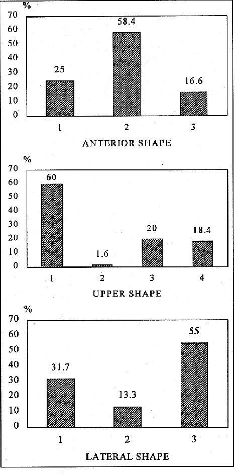
In the study of occlusal characteristics we observed that 36.6% of the
specimens presented teeth with cusp wear, 33.4% exhibited only facets of
wear, 16.7% marked wear or severe attrition, and 13.3% presented teeth
with no wear (Figure 2, top). Of the skulls studied, 66.6% presented normal
overbite, 23.4% had absent overbite, and 10% moderate overbite (Figure
2, middle). The Spee curve was moderate in 50% of the skulls, normal in
46.6% and marked in only 3.4% (Figure 2, bottom).
The correlations calculated between the morphologic, occlusal and dental
variables were statistically significant for the anterior, upper and lateral
condyle shape in relation to depth of the glenoid fossa, i.e., the depth
of the glenoid fossa was greater in skulls presenting a more rounded or
convex shape of this structure (Table 1). In contrast, there was no significant
correlation between the anterior, upper and lateral condyle shape and occlusal
characteristics such as Spee curve, overbite or tooth attrition. Another
significant correlation of the present study was the greater depth of the
glenoid fossa in skulls with normal overbite than in skulls with moderate
or absent overbite. No such correlation was detected between depth of the
glenoid fossa and Spee curve or tooth attrition (Table 1).
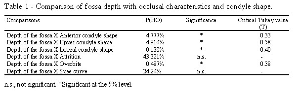
Figure
1 - Top, Percent distribution of the dry skull sample according to the
anterior shape of the mandibular condyle: 1, rounded or convex; 2, plane
or slightly convex; 3, shaped like an inverted "V". Middle, Percent distribution
of the dry skull sample according to the upper shape of the mandibular
condyle: 1, oblong; 2, rounded or oval; 3, laterally tapered, pear-shaped;
4, medially tapered, pear-shaped. Bottom, Percent distribution of the dry
skull sample according to the lateral shape of the mandibular condyle:
1, convex; 2, plane or slightly convex; 3, pointy or shaped like an inverted
"V".
Figure
2 - Top, Percent distribution of the dry skull sample according to tooth
wear or attrition: 0, no wear; 1, with facets of wear; 2, with cusp wear;
3, with marked wear or severe abrasion. Middle, Percent distribu-tion of
the dry skull sample according to overbite: 1, normal; 2, moderate; 3,
absent. Bottom, Percent distribution of the dry skull sample according
to Spee curve: 1, normal; 2, moderate; 3, marked.
Discussion
The skulls used in the present study were from individuals who died between
1957 and 1959. Considering that these skulls belong to contemporary human
beings, a more reliable evaluation of occlusal characteristics and of morphology
of the articular components would be expected. Many results of previous
studies, especially with respect to the characteristics of tooth wear or
attrition, are not consistent with the characteristics of human beings
of our times. Angel (1948) and Wedel et al. (1978) worked with medieval
material. Demirjian (1967) studied skulls from different collections. Seward
(1976) studied skulls of native Australians, while Granados (1979) evaluated
skulls of unknown origin, but there was evidence that many of them dated
back to the 19th century. Hinton (1981b) analyzed several samples ranging
from New World Aborigines to pre- and post-medieval individuals and even
white North Americans who lived in the industrial era.
Analysis of the results showed no statistically significant differences
between sexes for anteroposterior or mediolateral condyle dimensions or
depth of the glenoid fossa. However, the values obtained by Wedel et al.
(1978) and Hinton (1983) for mediolateral width were lower for women than
for men. Christiansen and Thompson (1990) also reported that the transverse
condylar dimension of normal adult joints was greater for men (19.6 mm)
than for women (17.7 mm). With respect to depth of the glenoid fossa, the
present results disagree with those reported by Demirjian (1967), who detected
highly significant differences between male (8.0 mm) and female (7.4 mm)
skulls. Furthermore, the dimensions detected by this investigator were
greater.
Considering condyle shape, the present results were similar to those
reported by Yale et al. (1966), who detected a slightly convex shape with
an anterior view in 59.4% of their skulls, and different from those obtained
by Öberg et al. (1971) and Wedel et al. (1978), who reported that
most of the condyles (51%) presented a convex shape in an anterior view.
As to the upper aspect, most of the condyles studied here (60%) presented
an oblong shape, confirming the results reported by Wedel et al. (1978).
Yale et al. (1961), however, detected this oblong shape only in slightly
convex condyles.
These contradictory data may be attributed to the fact that the investigators
cited worked with markedly different samples. Wedel et al. (1978), for
example, studied infant skulls (0 to 7 years), young skulls (7 to 14 years)
and juvenile skulls (14 to 20 years), in addition to adult skulls (older
than 20 years). In our investigation, we only studied skulls older than
18 years and especially in the 20-30 year range.
With respect to the occlusal characteristics, only 16.7% presented marked
tooth wear or severe tooth attrition, although this percentage was higher
in the samples studied by Demirjian (1967), Granados (1979) and Richards
(1987), among others.
In the present study there was a difference between sexes only in the
extent of tooth wear or attrition, which were greater in male specimens.
Demirjian (1967) also observed this tendency, with nineteen male skulls
presenting teeth with the greatest extent of attrition, whereas the female
skulls presented only initial levels of wear or attrition. According to
Hinton (1981a), this variation is probably the consequence of several factors,
among them a more intense dental overload in males.
The present results showed no significant correlation between condyle
shape and occlusal characteristics, in contrast to the results reported
in most of the previous studies. Mongini (1975), for example, stated that
the correlation between the morphologic appearance of the condyle and dental
attrition clearly shows that these two elements depend on the functional
pattern adopted by the masticatory apparatus. Granados (1979) and Richards
and Brown (1981) also detected severe alterations in the condyle of skulls
with teeth presenting marked wear. Richards (1987) and Wedel et al.(1978)
also reported the relationship between attrition and the rates of change
in condyle shape. This discordant result may possibly be attributed to
the difference between the samples analyzed in the present study and those
evaluated in previous studies. As mentioned earlier, these ancient collections
of dry skulls are from ancestors whose alimentary habits were quite different
from present ones, a fact that led to exaggerated tooth wear with an effect
on the morphology of the osseous components of the temporomandibular joint.
The amount of dental attrition detected in the skulls studied here was
much lower than in the previous studies.
The data obtained by Demirjian (1967) disagree with those obtained here
with respect to a greater depth of the glenoid fossa in skulls with a normal
overbite, since this investigator did not detect a significant correlation
between these variables. On the other hand, Angel (1948) observed that
small overbites are related to a plane glenoid fossa. These investigators
worked with specimens quite different from those used in the present study
in terms of morphologic characteristics and environmental variations such
as alimentation, diet and habits.
In agreement with the results reported by Demirjian (1967), we did not
detect a significant correlation between depth of the glenoid fossa and
extent of tooth wear or attrition. However, many investigators obtained
significant results, such as Hinton (1981 a, b, 1983) who detected an abrupt
reduction in the depth of the fossa in the presence of more severe levels
of molar wear or attrition. Similarly, Granados (1979) reported that loss
of cusp and anterior guide height occurring in the presence of marked attrition
was accompanied by severe resorption of the articular eminence, which became
shallower and, in some skulls, fully flattened.
The major reason for the divergences detected in the present study compared
to those reported by others is the fact that the samples used in the previous
studies consisted of specimens from ancestors whose alimentary habits provoked
masticatory stress due to ample occlusal forces and forces of repetitive
mastication, resulting in a bone morphology with its own characteristics.
Conclusions
The study of the morphology of the articular components and of the occlusal
characteristics of dry skulls demonstrated a significant correlation between
condyle shape and depth of the mandibular fossa, i.e., the depth of the
fossa was greater in skulls with more rounded condyles. No significant
correlation was observed between depth of the glenoid fossa and dental
attrition or Spee curve. However, there was a significant correlation between
deep glenoid fossae and skulls with normal overbite when compared to skulls
with moderate or deep overbite.
References
Angel JL: Factors in temporomandibular form. Am J Anat 83: 223-246, 1948
Angle EH: Classification of malocclusion. Dental Cosmos, 41: 248-264,
1899
Bell WE: Temporomandibular disorders: classification, diagnosis, management.
3rd ed. Year Book Medical, Chicago 1990
Christiansen EL, Thompson JR: Temporomandibular joint imaging. Mosby
Co., St. Louis 5-26, 1990
Demirjian A: A study of the morphology of the glenoid fossa. Nat Mus
Can Bull 206: 1-25, 1967
Dubrul EL: Sicher's Oral Anatomy, 7th ed., CV Mosby Co., St. Louis 1980
Gianelly AA, Ruben MP, Risinger R: Effect of experimentally altered
occlusal vertical dimension on temporomandibular joint. J Prosthet Dent,
24: 629-635, 1970
Granados JL: The influence of the loss of teeth and attrition on the
articular eminence. J Prosthet Dent 42: 78-85, 1979
Hinton RJ: Changes in articular eminence morphology with dental function.
Am J Phys Anthropol 54: 439-455, 1981a
Hinton RJ: Temporomandibular joint size adaptations in prehistoric Tennessee
Indians. Tenn Anthropol 6: 89-111, 1981b
Hinton RJ: Relationship between mandibular joint size and craniofacial
size in human groups. Arch Oral Biol 28: 37-43, 1983
Mongini F: Dental abrasion as a factor in remodeling of the mandibular
condyle. Acta Anat 92: 292-300, 1975
Öberg T, Carlsson GE, Fajers CM: The temporomandibular joint: a
morphologic study on a human autopsy material. Acta Odontol Scand 29: 349-384,
1971
Okeson JP: Management of temporomandibular disorders and occlusion.
2nd ed. CV Mosby, St. Louis 1-26, 1989
Richards LC: Temporomandibular joint: morphology in two Australian aboriginal
populations. J Dent Res 66: 1602-1607, 1987
Richards LC, Brown T: Dental attrition and degenerative arthritis of
the temporomandibular joint. J Oral Rehabil 8: 293-307, 1981
Seward ES: Tooth attrition and the temporomandibular joint. Angle Orthod,
46: 162-170, 1976
Wedel A, Carlsson GE, Sagne S: Temporomandibular joint morphology in
a medieval skull material. Swed Dent J 2: 177-187, 1978
Yale SH: Laminagraphic cephalometry in the analysis of mandibular condyle
morphology. Oral Surg Oral Med Oral Pathol 14: 793-805, 1961
Yale SH, Allison BD, Hauptfuehrer JD: An epidemiological assessment
of mandibular condyle morphology. Oral Surg Oral Med Oral Pathol 21: 169-177,
1966
Correspondence:Profa. Dra. Mírian Aiko Nakane Matsumoto,
Faculdade de Odontologia de Ribeirão Preto, USP, 14040-904 Ribeirão
Preto, SP, Brasil.
|


